Research
I have an interest in point-spread-function (PSF) engineering, super-resolution microscopy, light field microscopy, microfluidics and machine-learning-involved biomedical imaging techniques. I have two years' experience in wavefront-modulation-based PSF engineering using Bessel beam and Airy beam. I was also working on stochastic optical reconstruction microscopy (STORM) including cell culture, sample preparation, cell immunostaining, hardware control (lasers, AOTF, microscopic stages, cameras, etc.) data acquisition (using Hal-4000) and data post-processing and super-resolution image reconstruction. My current focus is light-field microscopy (LFM), especially Fourier light-field microscopy (FLFM). The Jia Lab are welcome to all potential collaborators who work in biological, biomedical, image processing and machine learning fields. The following are the detailed research experiences.
PhD research
Light-Field Flow Cytometry for High-Resolution, Volumetric, andMultiparametric 3D Single-Cell Analysis
Aug., 2021-Apr., 2023
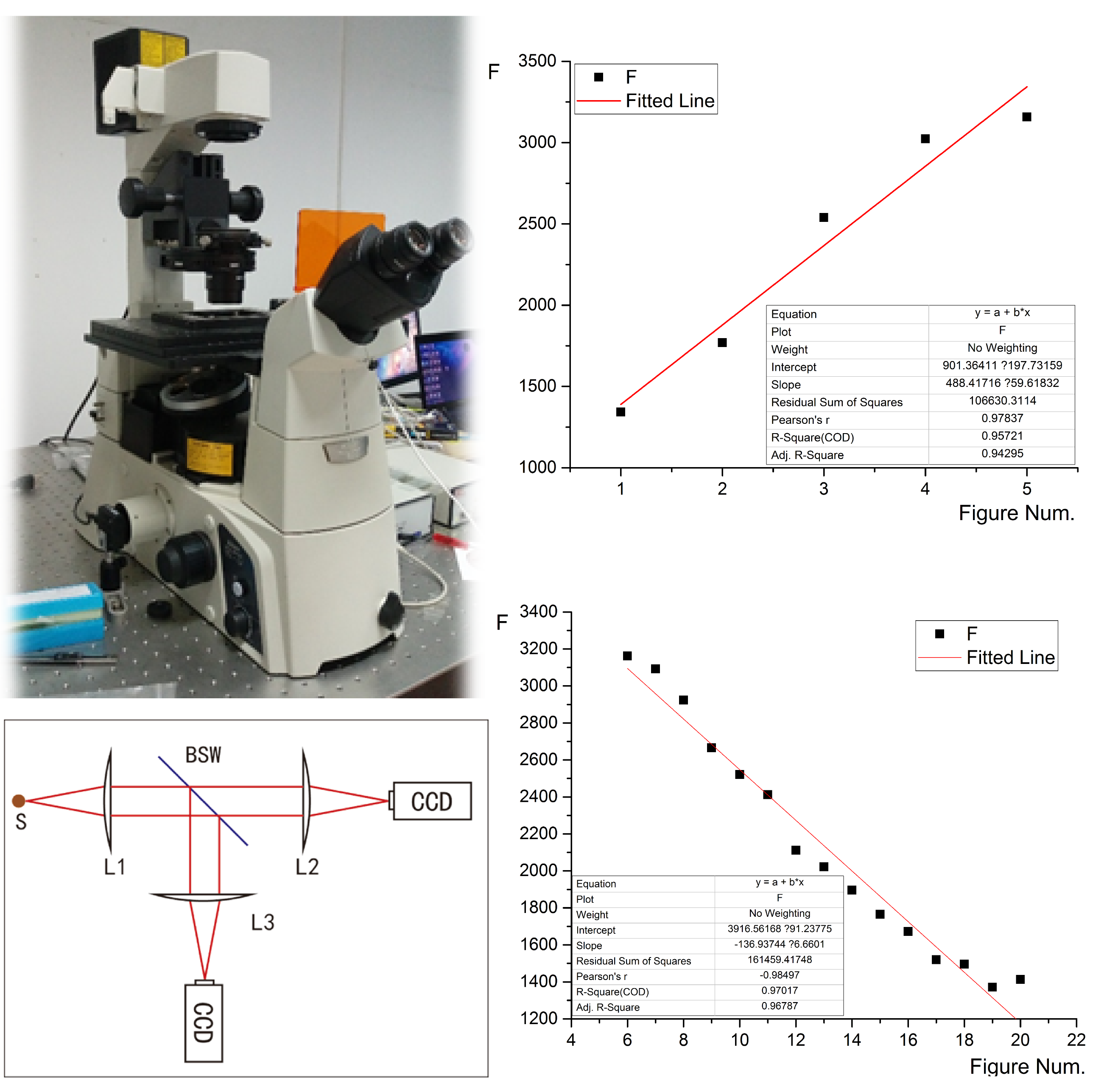
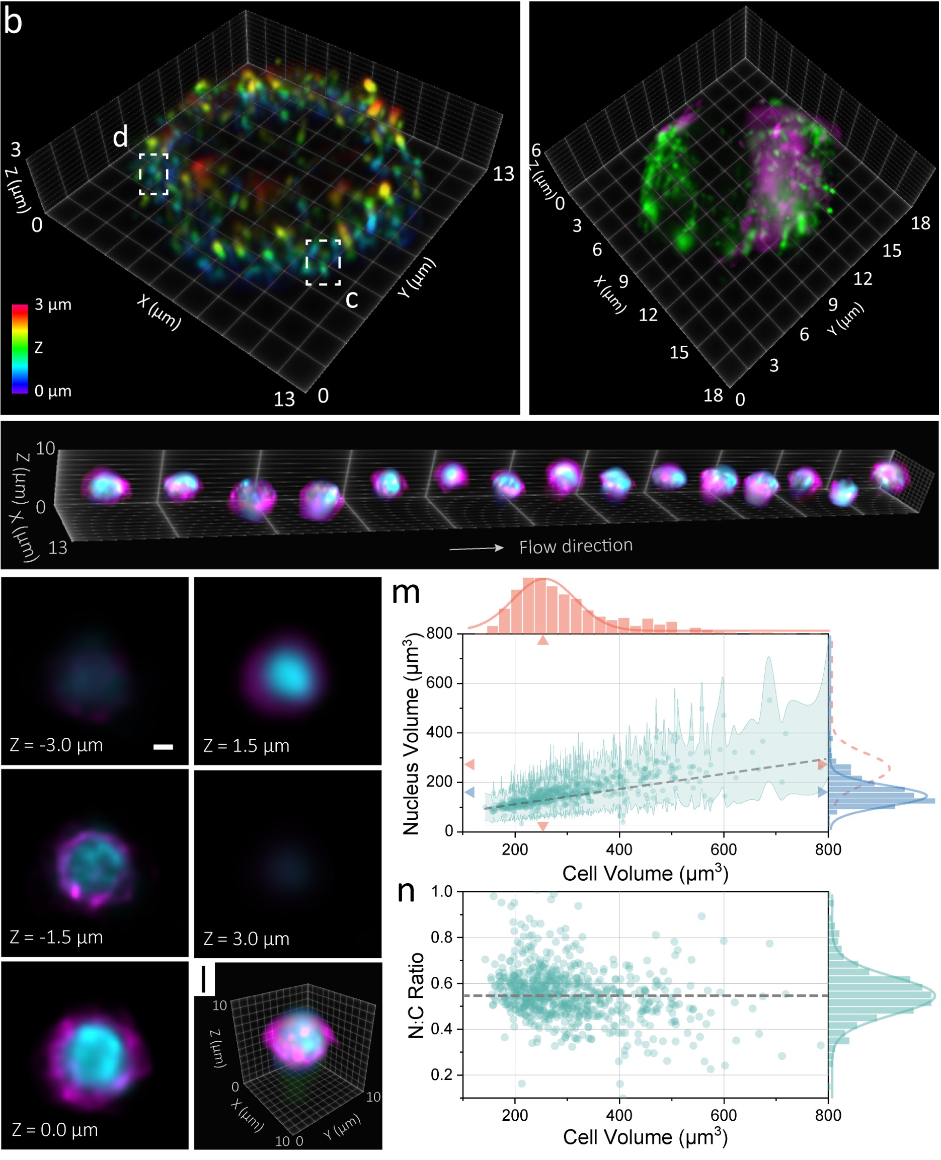
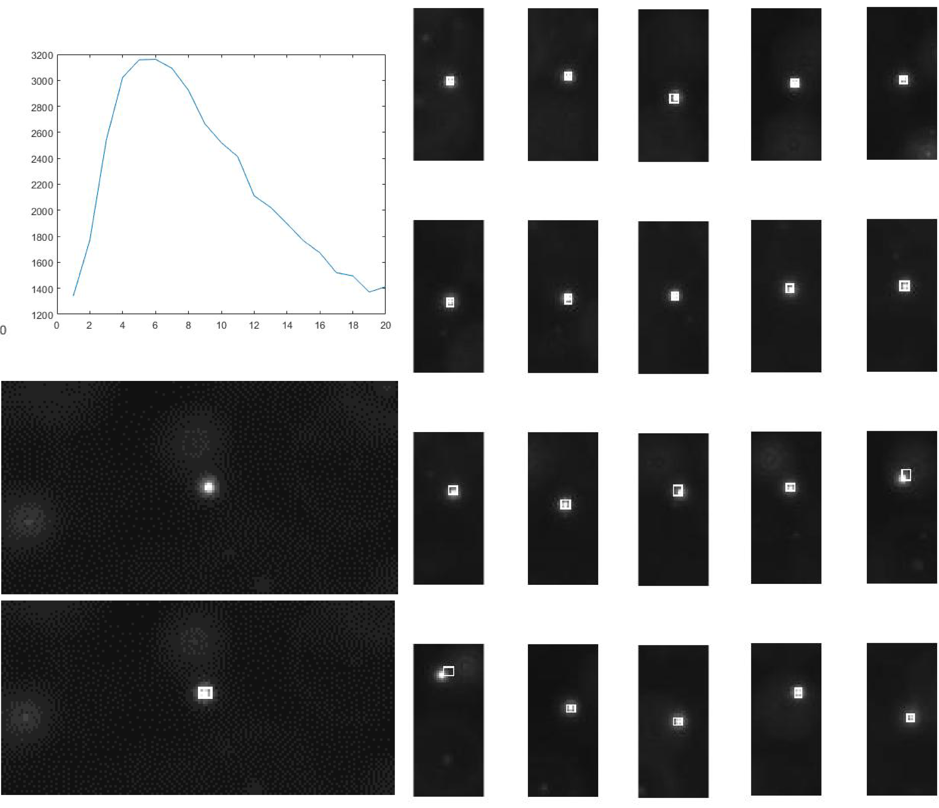
Imaging flow cytometry (IFC) combines flow cytometry and fluorescence microscopy to enable high-throughput, multiparametric single-cell analysis with rich spatial details. However, current IFC techniques remain limited in their ability to reveal subcellular information with a high 3D resolution, throughput, sensitivity, and instrumental simplicity. In this study, we introduce a light-field flow cytometer (LFC), an IFC system capable of high-content, single-shot, and multi-color acquisition of up to 5,750 cells per second with a near-diffraction-limited resolution of 400-600 nm in all three dimensions. The LFC system integrates optical, microfluidic, and computational strategies to facilitate the volumetric visualization of various 3D subcellular characteristics through convenient access to commonly used epi-fluorescence platforms. We demonstrate the effectiveness of LFC in assaying, analyzing, and enumerating intricate subcellular morphology, function, and heterogeneity using various phantoms and biological specimens. The advancement offered by the LFC system presents a promising methodological pathway for broad cell biological and translational discoveries, with the potential for widespread adoption in biomedical research.
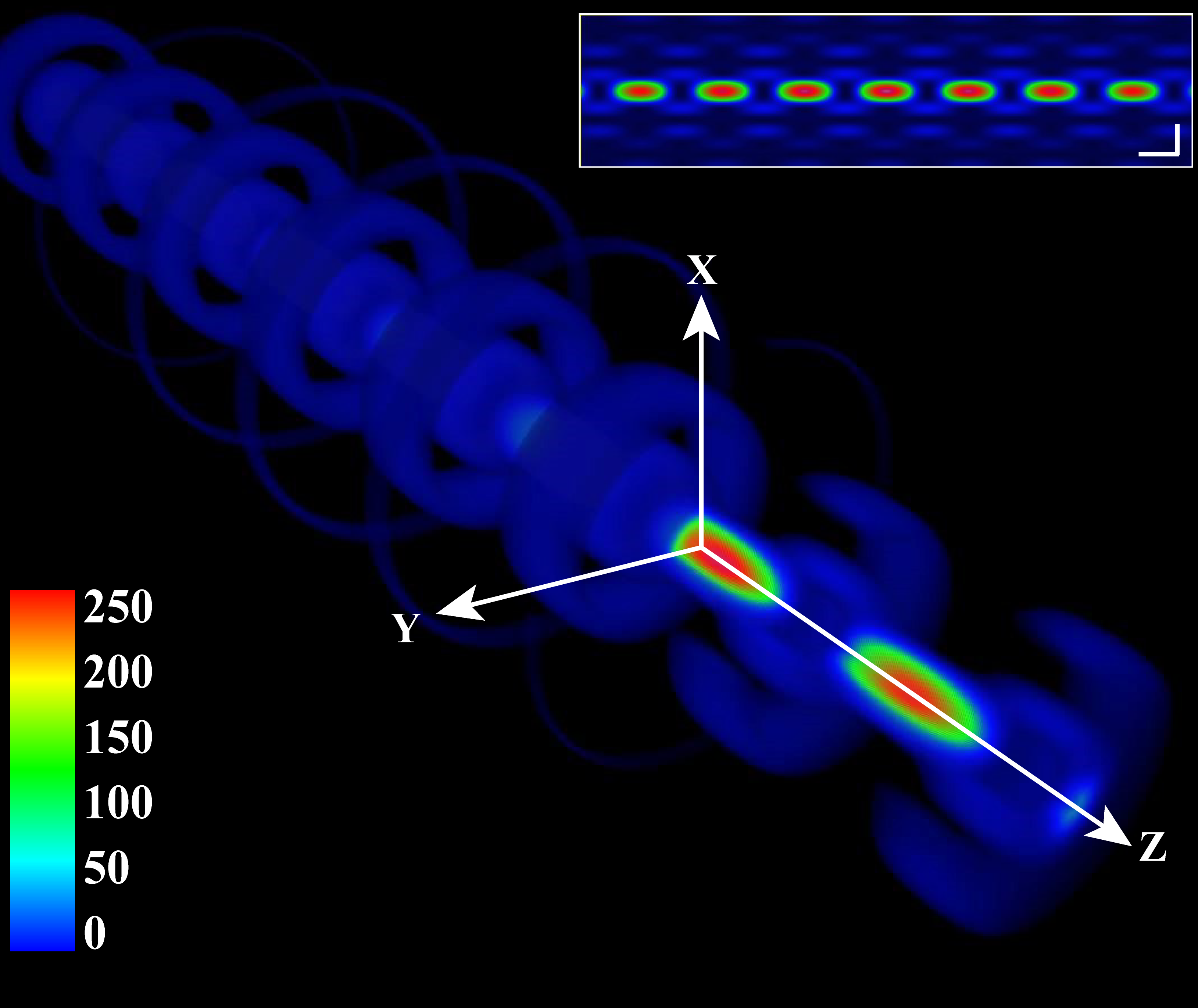
High-Resolution Fourier Light-Field Microscopy (HR-FLFM) for Volumetric Multi-Color Live-Cell Imaging
Oct., 2018-Apr., 2021
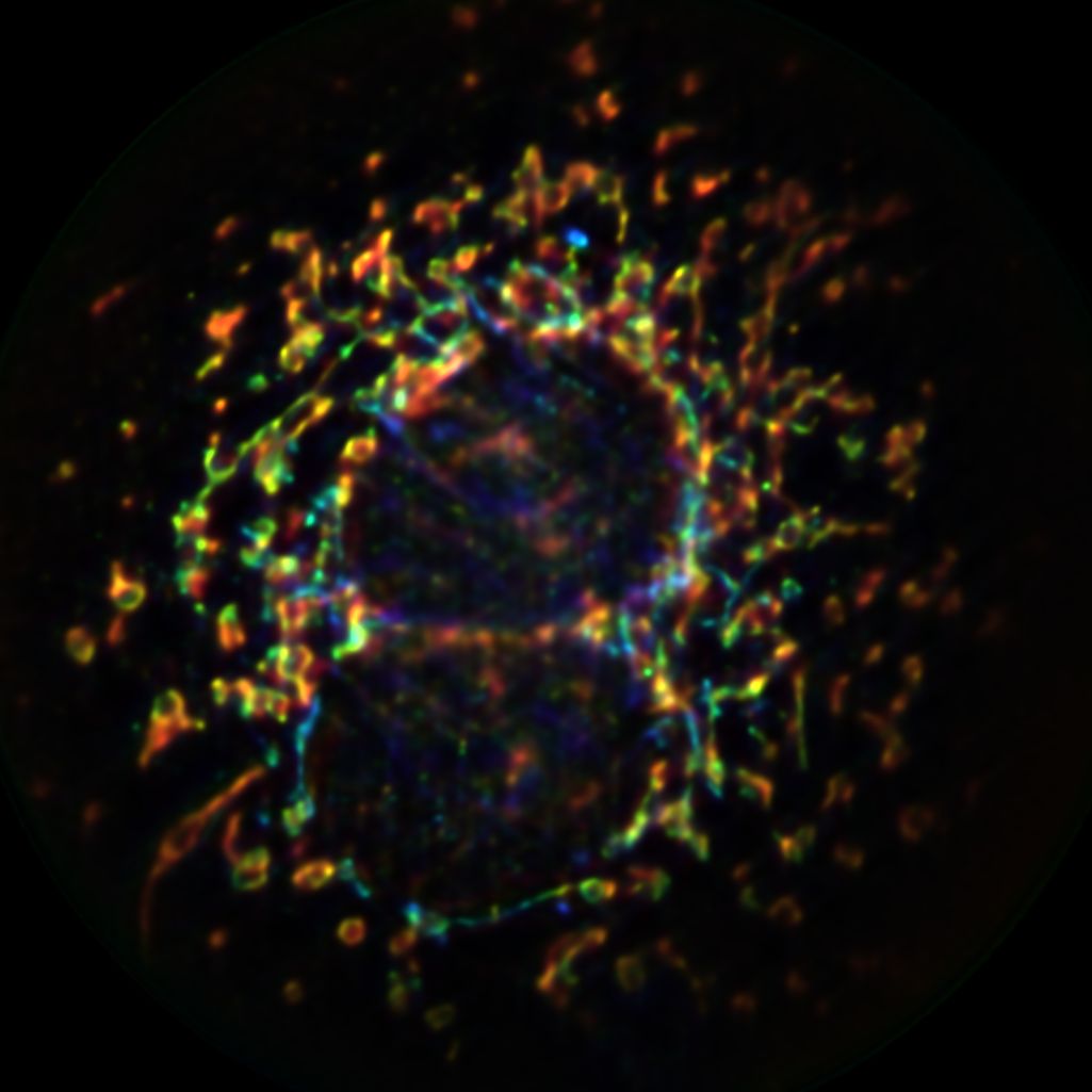
Volumetric interrogation of the organization and processes of intracellular organelles and molecules in cellular systems with a high spatiotemporal resolution is essential for understanding cell physiology, development, and pathology. Here, we report high-resolution Fourier light-field microscopy (HR-FLFM) for fast and volumetric live-cell imaging. HR-FLFM transforms conventional cell microscopy and enables exploration of less accessible spatiotemporal-limiting regimes for single-cell studies. The results present a near-diffraction-limited resolution in all three dimensions, a five-fold extended focal depth to several micrometers, and a scanning-free volume acquisition time up to milliseconds. The system demonstrates instrumentation accessibility, low photo damage for continuous observation, and high compatibility with general cell assays. We anticipate HR-FLFM to offer a promising methodological pathway for investigating a wide range of intracellular processes and functions with exquisite spatiotemporal contextual details.
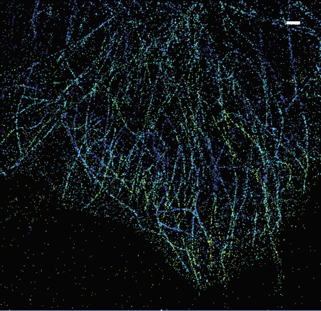
Stochastic Optical Reconstruction Microscopy (STORM)
Feb., 2018-present
Built up the optical system based on the Nikon Eclipse Ti-U fluorescent microscope. Developed a STORM controlling system based on the Hal-4000 source codes provided by Hazen Babcock. Performed both 2D STORM and 3D STORM imaging for both mitochondria and microtubules and achieved a 30-40 nm lateral resolution.
Depth-extended, high-resolution fluorescence microscopy: whole-cell imaging with double-ring phase (DRiP) modulation
Sep., 2016-Oct., 2018
We report a depth-extended, high-resolution fluorescence microscopy system based on interfering Bessel beams generated with double-ring phase (DRiP) modulation. The DRiP method effectively suppresses the Bessel side lobes, exhibiting a high resolution of the main lobe throughout a four- to five-fold improved depth of focus (DOF), compared to conventional wide-field microscopy. We showed both theoretically and experimentally the generation and propagation of a DRiP point-spread function (DRiP-PSF) of the imaging system. We further developed an approach for creating an axially-uniform DRiP-PSF and successfully demonstrated diffraction-limited, depth-extended imaging of cellular structures. We expect the DRiP method to contribute to the fast-developing field of non-diffracting-beam-enabled optical microscopy and be useful for various types of imaging modalities.
Undergraduate Research
Simultaneous Three-dimensional Tracking and Fluorescent Imaging of Microorganism Motility
May, 2014-Nov., 2015
Selected defocused microscopy to realize three-dimensional tracking from various approaches. Found appropriate functions in MATLAB to trace the living E. coli three-dimensionally based on the conclusion made in the project Exploration of Object-Image Relations under the Defocused Microscope. Designed the light path, constructed it and created programs in LabVIEW. Currently, adjusting the light path to match the configuration of the programs. Learning image processing and data analysis with MATLAB.
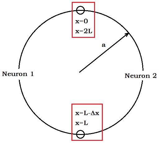
Simulation of action potential and magnetic field of two coupled neurons
Mar., 2015-May, 2015
Simplified the model of the action potential (AP) of two coupled neurons. Simulated the process of AP with MATLAB when external stimulations were injected into the two neurons. Figured out the characteristics of the surrounding magnetic field as the AP changed at different time.
Exploration of Object-Image Relations under the Defocused Microscope
Oct., 2014-Dec., 2014
Used the defocused microscopy to evaluate the effects of defocused imaging and analyzing the first-hand data. Confirmed the quadratic-linear model of the relationship between the defocused ring and the axial position of the bacteria.